Symptoms
We treat all types of brain tumors, including primary brain tumors (glioblastoma), brain and spine metastasis, pituitary tumors, meningiomas and skull base tumors (vestibular schwannomas). The signs and symptoms of tumors can vary significantly, depending on where the tumor is located, its size and how fast it is growing. Symptoms that could suggest a brain tumor include:
Headaches that become more intense and frequent
Weakness or paralysis in a body part, or one side of the body
Confusion or difficulty making decisions
Nausea or vomiting
Balance problems
Balance problems
Personality changes
Blurred vision, double vision or loss of peripheral vision
Speech difficulties
Seizures
Feeling exhausted
Hearing problems
Diagnosis
Brain tumors are sometimes discovered during imaging tests for another medical issue. But it can be challenging to diagnose a brain tumor and often involves several specialists working together. If you are experiencing brain tumor symptoms, your primary care doctor will start with a physical exam and a thorough exploration of your and your family’s medical history. A neurological exam may be used to look for issues with balance, hearing, vision, reflexes and mental health. If a brain tumor is suspected, additional tests will be performed, including:
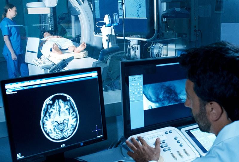
Magnetic resonance imaging (MRI) or computed tomography (CT) scans will be used to peer into your brain to look for signs of a tumor. They can pinpoint the exact location and size of a tumor.
If a tumor is discovered, your team will want to collect a sample so that it can be tested to identify the tumor type and to determine if it is cancerous. Often, your neurosurgeon will remove part or all of the tumor, if possible, during the biopsy. If the tumor is difficult to reach, the sample may be taken during a procedure that creates a small hole in your skull and a needle to remove tissue.
This may be done if your team suspects the tumor has crossed into the layers of tissue covering your brain. A small needle is used to collect cerebrospinal fluid from around your spine. It will be examined for cancer cells.
Treatments
We offer the latest in surgical and nonsurgical treatment options, with an emphasis on minimally invasive techniques. Among the procedures we provide:
This minimally invasive procedure allows your doctor to treat some tumors using an endoscope (a thin, rigid tube equipped with a tiny camera) inserted through the nose. The procedure, used for pituitary and skull base tumors, eliminates the need to open the skull and avoids brain retraction. It uses the natural nasal cavity to reach the tumor quickly and safely. That allows for faster recovery times and fewer complications.
One of the challenges of brain surgery is reaching the tumor without risking damage to surrounding areas. This minimally invasive cranial surgery uses tubular retractor systems to allow your neurosurgeon to reach tumors more easily in deeper sections of the brain. It uses a tube inserted through a small hole in the skull to reach the tumor. The tube displaces brain tissue (rather than cutting through it) on its way to the tumor. The surgeon then operates and removes tissue through the tube. Using endoscopes through these retractor systems, the surgeon can look within the tumor cavity to confirm complete and safe resection.
This minimally invasive procedure uses a thin laser fiber to destroy tumor tissue. Your neurosurgeon inserts the fiber through a small hole in your skull and guides it into position using magnetic resonance imaging (MRI). The procedure is effective against deep-seated tumors, and typically has a shorter hospital stay and faster recovery.
Sometimes referred to as internal radiation, this procedure involves placing radioactive material inside the body to kill a tumor. It allows your doctor to target a specific part of your body with a high dose of radiation. GammaTile is a collagen-based material that holds the radioactive material and limits its range – avoiding damage to healthy tissue.
Also called awake craniotomy, this is a surgery in which the patient is kept awake, but comfortable with anesthesia and numbing medications. This type of surgery is particularly advantageous in cases where the tumor is located near functional brain tissue. Your neurosurgeon can get immediate feedback from you during the surgery, reducing the risk of neurological complications.
This minimally invasive procedure is designed to shut off the blood supply to a tumor. To do this, your surgeon will make a tiny incision in your groin or forearm and thread a catheter – guided by advanced imaging – to the site. Special material will then be injected, closing off the blood flow. This procedure tends to work better with tumors with a prominent blood supply. Sometimes this procedure is done before surgery, as it decreases the blood loss during surgery.
For some procedures, we employ a robotic surgical assistant that increases surgical speed and accuracy. ROSA still requires the expertise of a neurosurgeon, but it allows many procedures to be done in a minimally invasive manner, reducing recovery time and potential complications.
Among the cutting edge pre-surgical technologies we offer is Quicktome, a system designed to precisely map a patient’s unique brain network. The system starts with an MRI scan of your brain and applies algorithms that analyze millions of data points to build your brain map. Your brain network controls a wide range of functions, including language, movement and decision making. Having this map reduces the risk of losing brain function during your surgery.
Orlando Health Cancer Institute, partnered with the Neuroscience Institute is the only center in Orlando to offer this treatment, which uses proton beams to treat some cancers. While X-ray radiation has long been used to treat cancer, this newer technology allows your doctor to precisely focus on the target zone, potentially resulting in fewer unwanted side effects.
This cutting-edge procedure, which will be available next year, combines surgery with advanced imaging. Traditionally, a patient would receive an MRI scan before surgery and again afterward to confirm the procedure went as planned. But with intraoperative MRI, additional scans can be made while the procedure is happening to rule out residual that can still be safely taken out.
Our team participates in clinical trials that can offer our patients early access to promising treatments, therapies and surgeries.
Brain (Pituitary) Tumors Treatment at Orlando Health Neuroscience Institute
Our neuro-oncology team has access to the most advanced treatment options for brain and spinal tumors. Your treatment options are determined by a tumor’s location, behavior and cell type. With all types of brain and spine tumors, skill and technology are vital to treatment outcomes. The Orlando Health Cancer Institute provides both.